


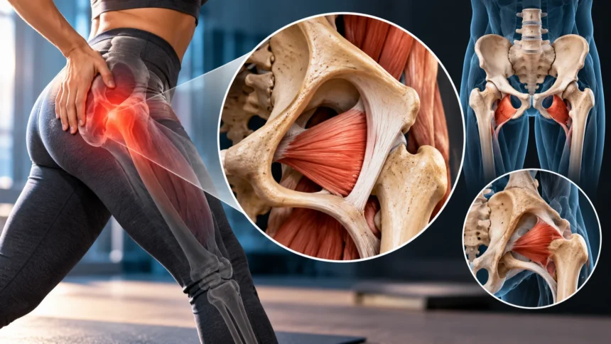
What Is the Obturator Muscle? Anatomy, Function & Key Facts
The obturator muscle plays an important role in hip movement, pelvic stability, and lower-body coordination. Although these muscles are not as widely discussed as the glutes or quadriceps, they help control how the thigh moves within the hip joint. Understanding obturator muscle anatomy can provide valuable insight into everyday movements, exercise performance, and certain causes of hip or pelvic discomfort.
The term obturator muscle generally refers to two separate muscles: the obturator internus and obturator externus. Both are located around the pelvis and contribute to rotating and stabilizing the hip, but they differ in their anatomical positions, attachments, and additional functions. They work alongside other deep hip muscles to support controlled movement during walking, standing, and physical activity.
What Is the Obturator Muscle?
The obturator muscles are a pair of deep muscles associated with the pelvic region and hip joint. They include the obturator internus and obturator externus, which are positioned on different sides of the obturator membrane. Both muscles help control rotation of the thigh and contribute to the stability of the hip during movement.
These muscles originate from structures surrounding the obturator foramen, a large opening within each hip bone. The obturator membrane covers most of this opening and provides attachment surfaces for nearby muscles. Although the two obturator muscles have different anatomical pathways, both connect to the femur and influence the position of the thigh.
The obturator muscles are especially important because hip movement requires cooperation between large and small muscle groups. While the gluteal muscles generate substantial force, deeper muscles help guide and stabilize the joint. This combination allows the lower body to perform coordinated movements without relying exclusively on larger muscles for stability and control.
Obturator Muscle Anatomy and Location
Obturator muscle anatomy begins with the structure of the pelvis, which contains the hip bones, sacrum, and surrounding connective tissues. The obturator foramen is formed by portions of the pubis and ischium. The obturator internus occupies the internal surface surrounding this opening, while the obturator externus lies on its outer surface.
The obturator internus follows a distinctive anatomical pathway because its tendon exits the pelvis through the lesser sciatic foramen. It then changes direction around the ischium before attaching to the upper femur. This arrangement allows the muscle to influence hip rotation while also contributing to the mechanical support of the hip joint.
The obturator externus follows a shorter pathway from the outer pelvic surface toward the femur. It passes behind the femoral neck before reaching its attachment near the greater trochanter. Its deep location makes it difficult to identify through surface examination alone, which is why detailed anatomical knowledge is useful during clinical assessment.
Obturator Internus Muscle: Structure and Attachments
The obturator internus is a deep hip muscle that originates from the pelvic surface of the obturator membrane and surrounding pelvic bones. Its muscular fibers converge toward a tendon that travels through the lesser sciatic foramen. The tendon then turns sharply around the lesser sciatic notch before approaching the femur.
The muscle inserts onto the medial surface of the greater trochanter, near the trochanteric fossa. Along its course, the tendon is supported by surrounding connective tissues and a bursa that reduces friction as it changes direction. This distinctive arrangement allows the muscle to contribute to controlled rotation and stabilization of the hip.
The obturator internus is commonly grouped with the gemellus superior and gemellus inferior muscles because their tendons are closely associated. Together, they form part of the deep hip rotator system. Their coordinated activity helps maintain joint positioning while supporting rotational movements that occur during walking, changing direction, and other daily activities.
Obturator Externus Muscle: Structure and Attachments
The obturator externus is a flattened, triangular muscle located on the outer surface of the obturator membrane and adjacent pelvic bones. It originates from the external surfaces of the pubis and ischium surrounding the obturator foramen. Its fibers travel backward and outward toward the proximal portion of the femur.
The muscle’s tendon passes beneath the femoral neck and inserts into the trochanteric fossa of the femur. This position gives the obturator externus a useful mechanical advantage for controlling external rotation. It also allows the muscle to contribute to joint stability while the hip experiences changing loads during movement.
Although the obturator externus is relatively small, its location makes it important for coordinated hip mechanics. It works alongside other deep rotators to guide the femoral head within the hip socket. This contribution becomes especially relevant during activities that require balance, directional changes, and precise control of the lower limbs.
Obturator Internus vs. Obturator Externus
The main difference between the obturator internus and obturator externus is their location relative to the obturator membrane. The internus originates primarily inside the pelvis, whereas the externus originates on the external pelvic surface. Both influence hip rotation, but their separate anatomical pathways create differences in their mechanical actions.
The obturator internus primarily contributes to external rotation when the hip is extended and can assist abduction when the hip is flexed. The obturator externus also helps externally rotate the thigh and stabilize the hip joint. Their exact mechanical contributions can vary depending on joint position and the activity being performed.
Another important difference involves their nerve supply and relationships with surrounding structures. The obturator internus receives innervation from the nerve to obturator internus, while the obturator externus is supplied by the obturator nerve. Recognizing these distinctions helps healthcare professionals interpret muscular symptoms and understand potential nerve-related movement problems.
What Is the Main Function of the Obturator Muscle?
The primary function of both obturator muscles is to support external rotation of the hip. External rotation occurs when the thigh rotates outward relative to the pelvis. This movement contributes to activities such as turning the leg, changing walking direction, and positioning the lower body during certain exercise movements.
The obturator muscles also help stabilize the femoral head within the acetabulum, commonly called the hip socket. This stabilizing action supports joint alignment and coordinated movement during weight-bearing activities. Their role becomes particularly important when the hip moves through different angles or responds to changing physical demands.
The obturator internus has an additional function because it can assist with hip abduction when the joint is flexed. Abduction involves moving the thigh away from the body’s midline. Together, these movements demonstrate how small hip muscles contribute to flexibility, balance, and controlled motion rather than simply producing large amounts of force.
How the Obturator Muscles Support Hip Stability
Hip stability depends on the coordinated activity of bones, ligaments, joint structures, and surrounding muscles. The obturator muscles contribute by helping control the position and rotation of the femoral head. Their deep attachments allow them to support the joint while larger muscles generate the force needed for walking and exercise.
During standing and walking, the hip must remain stable while supporting body weight and responding to movement. The obturator muscles work with the gluteal muscles and other deep rotators to maintain coordinated joint mechanics. Their activity changes according to hip position, movement speed, and the demands placed on the lower body.
Healthy hip stabilization is not simply about muscle strength. It also requires adequate mobility, efficient muscle coordination, and appropriate movement patterns. When these elements work together, the hip can respond effectively to everyday activities such as climbing stairs, changing direction, and maintaining balance on uneven surfaces.
Obturator Muscle Nerve Supply and Blood Supply
The obturator internus receives its nerve supply from the nerve to obturator internus, which usually arises from spinal nerve roots L5, S1, and S2. This nerve also supplies the superior gemellus muscle. Normal nerve function allows the obturator internus to contract appropriately and participate in coordinated hip movement.
The obturator externus receives its nerve supply from the posterior division of the obturator nerve, which generally contains fibers from L2, L3, and L4. The obturator nerve also contributes to the function of several thigh adductor muscles. Because of these relationships, nerve problems may affect multiple muscles rather than the obturator externus alone.
Blood reaches these muscles through branches of nearby pelvic and thigh arteries. The obturator internus receives contributions from vessels including the internal pudendal and gluteal arterial systems, while the obturator externus receives blood from obturator and circumflex femoral branches. Adequate circulation supports muscle function, tissue maintenance, and recovery from normal physical activity.
Obturator Muscle Pain: Common Causes
Obturator muscle discomfort can develop from muscular overload, repetitive movement, or problems involving nearby joints and tissues. Activities requiring frequent hip rotation may place additional demands on the deep hip muscles. However, discomfort in this region is not automatically caused by the obturator muscles because several structures can produce similar symptoms.
The obturator internus may be associated with certain forms of deep buttock or pelvic discomfort. Increased muscle sensitivity, altered muscular coordination, and surrounding tissue irritation can contribute to pain in some individuals. Conditions involving the hip joint, pelvic floor, nerves, or lower back can also produce overlapping symptoms that require careful evaluation.
Obturator externus injuries are relatively uncommon but may occur during demanding athletic movements or sudden changes in hip position. Possible problems include muscular strains and irritation near its attachments. Persistent symptoms should not be diagnosed from location alone, because a qualified healthcare professional may need to assess several possible causes.
Symptoms of Obturator Muscle Dysfunction
Symptoms associated with obturator muscle problems can vary according to the affected muscle and underlying condition. Some individuals experience deep hip discomfort, buttock pain, or pain during particular rotational movements. Others notice reduced movement tolerance or discomfort after prolonged activity, although these symptoms are not specific to obturator muscle dysfunction.
Obturator internus involvement may be considered in some cases of persistent pelvic pain or discomfort during sitting. The muscle’s relationship with deep pelvic structures makes clinical evaluation especially important when symptoms are difficult to localize. Pelvic floor muscle dysfunction and other conditions may coexist, making professional assessment more useful than relying on a single symptom.
Movement limitations can occasionally develop when discomfort causes protective muscular guarding or changes in normal coordination. However, weakness, numbness, radiating pain, or significant loss of function may indicate another condition. Sudden severe pain, inability to bear weight, or new neurological symptoms require prompt medical assessment rather than self-directed muscle treatment.
How Obturator Muscle Problems Are Diagnosed
Diagnosing obturator muscle problems begins with a detailed medical history and physical examination. A healthcare professional may ask about symptom location, activity levels, previous injuries, and movements that increase discomfort. This information helps determine whether symptoms may originate from the hip muscles, joints, pelvic structures, or surrounding nerves.
The physical examination may include assessing hip range of motion, muscular strength, walking patterns, and responses to specific movements. Clinicians sometimes evaluate discomfort during resisted hip rotation or other controlled tests. These findings must be interpreted alongside the broader examination because no single movement test definitively identifies every obturator-related condition.
Imaging studies may be recommended when clinicians suspect a significant injury or another underlying problem. Magnetic resonance imaging can sometimes reveal muscular injury, inflammation, or abnormalities in nearby structures. Imaging is not necessary for every case, and treatment decisions generally depend on the overall clinical picture rather than one isolated finding.
Obturator Muscle Exercises and Mobility
Gentle hip mobility exercises can support comfortable movement and help maintain flexibility around the pelvis. Controlled hip rotation, easy walking, and comfortable range-of-motion activities may be appropriate for individuals without significant symptoms. Exercise choices should reflect personal ability, existing conditions, and whether particular movements increase discomfort.
Hip strengthening programs often include exercises that target the gluteal muscles and improve lower-body control. Bridges, supported balance activities, and carefully selected hip rotation movements can help develop broader muscular coordination. These exercises do not isolate the obturator muscles completely, but they may support the overall hip system in which these muscles operate.
Stretching and strengthening should remain comfortable rather than forcing the hip into painful positions. Individuals experiencing persistent pelvic discomfort, deep hip pain, or suspected muscle injury may benefit from guidance provided by a qualified physical therapist. A personalized rehabilitation plan can address mobility, strength, movement habits, and any contributing conditions identified during assessment.
Can the Obturator Muscle Affect Pelvic Floor Function?
The obturator internus has an important anatomical relationship with the pelvic floor through the obturator fascia and surrounding connective tissues. Portions of this fascia contribute to the attachment and support of pelvic floor structures. This relationship helps explain why clinicians sometimes consider the obturator internus when evaluating certain pelvic pain and muscular coordination problems.
The pelvic floor consists of muscles and connective tissues that support pelvic organs and contribute to bladder, bowel, and other physiological functions. The obturator internus is not itself a pelvic floor muscle, but it lies close to this muscular system. Its condition may influence regional comfort and movement in some people.
Not every pelvic floor symptom is related to the obturator internus, and the connection should not be assumed without assessment. Pelvic discomfort can have many muscular, neurological, urinary, gastrointestinal, and other causes. A qualified healthcare professional or pelvic health physical therapist can help identify relevant factors and recommend appropriate management strategies.
Treatment and Recovery for Obturator Muscle Problems
Treatment for suspected obturator muscle dysfunction depends on the underlying cause and the severity of symptoms. Mild muscular discomfort may improve with temporary activity modification, comfortable movement, and gradual return to usual activities. However, persistent or worsening pain requires assessment to rule out joint injuries, nerve conditions, or other sources of discomfort.
Physical therapy may involve progressive strengthening, mobility exercises, movement retraining, and education about managing activity levels. When pelvic floor symptoms are present, treatment may also address muscular coordination and relaxation rather than focusing exclusively on strengthening. The most appropriate approach depends on examination findings, symptom patterns, and individual functional goals.
Recovery time varies because different conditions can affect the obturator region. Minor muscular irritation may settle relatively quickly, while more complex injuries or persistent pelvic pain can require longer management. Individuals should follow professional guidance, avoid repeatedly provoking significant symptoms, and seek reassessment when progress does not match the expected recovery pattern.
Conclusion
The obturator muscle system includes the obturator internus and obturator externus, two deep muscles that contribute to hip rotation and stability. Although they share several functions, their anatomical positions, attachments, and nerve supplies differ. Understanding these differences provides useful insight into how the pelvis and hip work together during everyday movement.
These muscles support coordinated lower-body activity and have important relationships with nearby joints, nerves, and pelvic structures. Problems affecting the obturator region may contribute to discomfort, but symptoms often overlap with other conditions. Accurate assessment is therefore essential when identifying the source of persistent hip or pelvic pain.
Maintaining healthy hip movement involves balanced muscular strength, comfortable mobility, and appropriate physical activity. Understanding obturator muscle anatomy can help individuals make informed decisions about exercise and recognize when professional guidance may be necessary. With proper evaluation and individualized care, many movement-related problems can be managed effectively.
Frequently Asked Questions
What is the obturator muscle responsible for?
The obturator muscles primarily help rotate the thigh outward and stabilize the hip joint. The obturator internus can also assist with moving the thigh outward when the hip is flexed.
Where is the obturator muscle located?
The obturator internus lies along the inner surface of the pelvis, while the obturator externus lies on the outer surface. Both originate around the obturator foramen and attach to the upper femur.
What causes obturator muscle pain?
Possible causes include muscular strain, repetitive loading, local tissue irritation, or problems involving nearby structures. Because deep hip and pelvic pain have several possible causes, persistent symptoms should be professionally evaluated.
Can obturator muscle problems cause pelvic pain?
The obturator internus may contribute to certain pelvic pain conditions because of its close anatomical relationship with pelvic structures. However, pelvic pain has many possible causes and requires appropriate clinical assessment.
How do you strengthen the obturator muscles?
Controlled hip rotation exercises and broader hip strengthening activities may help support obturator muscle function. A physical therapist can recommend suitable exercises based on individual mobility, strength, symptoms, and rehabilitation needs.